查看: 95129|回复: 16

胸腺病变:图文综述

[复制链接]

823

主题

368

回帖

292

日志

荣誉会员

积分
6487
QQ
发表于 2021-5-31 10:41:38 | 显示全部楼层 |阅读模式
本帖最后由 hyc3140 于 2021-6-1 10:50 编辑

胸腺病变:图文综述
Thymic lesions: a pictorial review
复习正常胸腺解剖。
探讨胸腺良、恶性及罕见病变在不同诊断技术中的影像学表现。
回顾可以诊断多发性胸腺病变的影像学参数。
展示鉴别诊断。

To review the normal thymic anatomy.
To describe the imaging features of benign, malignant, and rare thymic lesions in the different diagnostic techniques.
To review the imaging parameters that allow the diagnosis of multiple thymic lesions.
Expose the differential diagnosis.

背景
胸腺是免疫系统的淋巴器官,被认为是T淋巴细胞成熟的主要部位[1,2]。它从第3和第4鳃袋两侧出现,在妊娠第6周开始发育[1-3]。随后,组织向纵隔和尾部迁移到前纵隔。随后,淋巴细胞从肝脏和骨髓迁移到胸腺,导致腺体分叶状,随后,胸腺分化为皮质和髓质[2]。由于迁移,异位胸腺组织或病变可能发生在沿着这一束的胸腺导管(图1)。

胸腺在青春期达到最大重量。成年后,上皮细胞被脂肪组织取代[1]。

识别正常胸腺并将其与良性或恶性的病理状态区分开来是至关重要的。此外,由于它位于前纵隔,了解其他前纵隔肿块(例如脂肪瘤、脂肪肉瘤、淋巴管瘤、异位甲状腺、甲状旁腺病变和结节病)的鉴别诊断也很重要[1]。

Background
The thymus is a lymphoid organ of the immune system, and it is considered the primary site of T-lymphocyte maturation [1,2]. It emerges bilaterally from the 3rd and 4th branchial pouches, and the development begins in the 6th gestational week [1-3]. Later, the tissue migrates medially and caudally to the anterior mediastinum. Following that, there is a migration of the lymphoid cells from the liver and bone marrow to the thymus, leading to lobulation of the gland, and subsequently, the thymus differentiates into a cortex and medulla [2]. Due to the migration, ectopic thymic tissue or lesions may occur anyway along this tract of the thymopharyngeal duct (figure 1).

The thymus reaches the maximum weight in puberty. At adult age, the epithelial cells are replaced by adipose tissue [1].

It is crucial to recognize the normal thymus and distinguish it from pathological conditions, either benign or malignant. Also, since it is located at the anterior mediastinum it is important to understand the differential diagnosis of other anterior mediastinal masses (ex: lipoma, liposarcoma, lymphangioma, ectopic thyroid, parathyroid lesions, and sarcoidosis) [1].
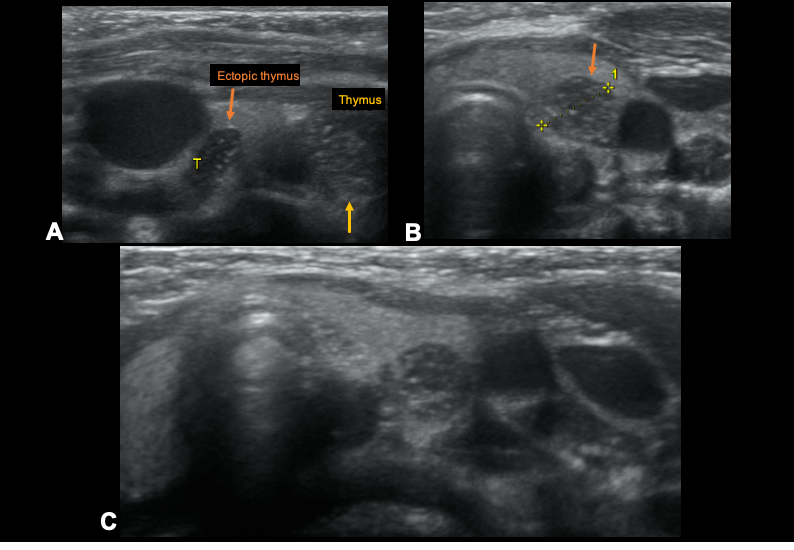
901484.jpg
图1:3岁患者颈部超声显示甲状腺左叶异位胸腺。低回声结节伴细的高回声斑点,显示与胸腺相同的回声结构,与异位胸腺相容。异位胸腺(橙色箭头)。胸腺(黄色箭头)。
Fig 1: Cervical ultrasound of a 3-year-old patient showing an ectopic thymus in the left lobe of the thyroid. Hypoechogenic nodule with fine hyperechogenic speckles demonstrating an echo-structure identical to the thymus, compatible with an ectopic thymus. Ectopic thymus (orange arrow). Thymus (yellow arrow).


1.正常解剖和组织学
正常胸腺通常位于前纵隔。在计算机断层扫描(CT)中,它通常被视为一个双叶三角形结构,婴儿较大,但成年后逐渐退化(图2)[1,4]。胸腺覆盖心包、主动脉弓、左无名静脉和气管[4]。据描述,与女性相比,年轻成年男性的脂肪萎缩发生得更快[5]。
组织学上,胸腺组织成多个小叶,排列成一个内髓质,内髓质含有成熟的淋巴细胞和旋涡状的梭形上皮细胞,这些细胞形成具有角化核心的Hassall小体;外皮质由未成熟的T淋巴细胞和胸腺上皮细胞组成[2]。
Fig 2 Non-contrast CT in a young patient, showing the normal configuration of th.gif
图2:年轻患者的平扫CT,显示胸腺的正常形态,本例为箭头形状(B)。
Fig 2: Non-contrast CT in a young patient, showing the normal configuration of the thymus, in this case with an arrow-head shape (B).

1. NORMAL ANATOMY AND HISTOLOGY
The normal thymus is normally located in the anterior mediastinum. At computed tomography (CT) it is usually visualized as a bi-lobed triangular structure, that is larger in infants but gradually involutes in adulthood (figure 2) [1,4]. The thymus overlies the pericardium, aortic arch, left innominate vein, and trachea [4]. It has been described that fatty atrophy occurs more rapidly in young adult men compared to women [5].

Histologically, the thymus is organized in multiple lobules arranged into an inner medulla which contains mature lymphocytes and whorls of spindle-shaped epithelial cells, which form Hassall corpuscles with keratinized cores; and an outer cortex which is composed of immature T-lymphocytes and thymic epithelial cells [2].
希望疫情早日结束!
回复 关闭延时

使用道具 举报

823

主题

368

回帖

292

日志

荣誉会员

积分
6487
QQ
发表于 2021-5-31 10:45:15 | 显示全部楼层
2.胸腺病变
胸腺病变可分为良性和恶性,但通常以胸腺瘤为主(图3-表1)。影像学在胸腺病变的鉴别、分期、手术指导和随访中起着重要作用。
901486.jpg
图3:表1。胸腺病变分为良性、恶性和罕见。
Fig 3: Table 1. Thymic pathologies grouped into benign, malignant, and rare.

2. THYMIC PATHOLOGIES
Thymic pathologies can be divided into benign and malignant, but they are generally dominated by thymomas (figure 3 - table 1). Imaging plays an important role in the identification, staging, surgical guidance, and follow-up of thymic lesions.


希望疫情早日结束!
回复 支持 反对

使用道具 举报

823

主题

368

回帖

292

日志

荣誉会员

积分
6487
QQ
发表于 2021-5-31 10:52:20 | 显示全部楼层
本帖最后由 hyc3140 于 2021-6-4 16:15 编辑

一。良性病变
A) 胸腺囊肿
胸腺囊肿占所有纵隔肿块的1-3%,可为先天性或后天性[1]。先天性囊肿通常是单房的薄壁囊肿。同时,获得性囊肿通常为多房性,壁厚,与重症肌无力、干燥综合征、狼疮、再生障碍性贫血、淋巴瘤和放射治疗有关(图4)。此外,后天性囊肿可能与胸腺恶性肿瘤有关。已有研究表明,MRI能更准确地鉴别胸腺囊肿和实质性病变[2]。

I. BENIGN LESIONS
A) THYMIC CYSTS
Thymic cysts represent 1-3% of all mediastinal masses and can be congenital or acquired [1]. Congenital cysts are usually unilocular with a thin wall. Meanwhile, the acquired cysts are generally multilocular with a thick wall and are associated with myasthenia gravis, Sjogren syndrome, lupus, aplastic anemia, lymphoma, and radiation therapy (figure 4). Furthermore, the latter cysts can be associated with thymic malignancy. It has been shown that MRI is more accurate in distinguishing thymic cyst from solid thymic lesions [2].

901528.jpg
图4:男,37岁。胸部正面X线片显示右纵隔轮廓异常(箭头)。增强CT(轴向B,矢状C)显示右前纵膈,液体密度肿块,无内强化,显示部分顶叶钙化。组织学显示胸腺组织有实性和囊性区域。
Fig 4: Male, 37 years old. A- Frontal X-ray of the chest reveals an abnormal right mediastinal contour (arrow). Contrast-enhanced CT (axial – B, sagittal-C), shows in the right anterior mediastinum, a fluid-attenuation mass without inner enhancement, showing some parietal calcifications. Histology revealed thymic tissue with solid and cystic areas.

B) 胸腺增生
胸腺增生有两种不同的组织学类型:
真性胸腺增生:组织学正常,是由于压力反弹(放疗或化疗、类固醇治疗、肺炎、手术或烧伤后)引起的。
淋巴滤泡增生:与自身免疫性疾病相关的腺体髓质淋巴滤泡数量增加[1,2]。
两者都被定义为胸腺体积对称性增大,使得它们在影像学上无法区分[2]。然而,由于其影像学表现,有可能将其与肿瘤区分开。它们通常表现为弥漫性和对称性肿大,平滑的轮廓和散布的脂肪和软组织成分,保存的脂肪平面和正常的血管(图5)。此外,由于存在微观脂肪,MRI在检测它们时具有很高的灵敏度,在T1加权异相图像上显示信号强度下降[2,4]。通常,胸腺上皮性肿瘤在磁共振成像中没有表现出这些特征,这使得这一发现有助于区分这些特征。

B) THYMIC HYPERPLASIA
There are two distinct histologic types of thymic hyperplasia:
    True thymic hyperplasia: normal histology and happens due to a rebound from stress (after radiation or chemotherapy, steroid treatment, pneumonia, surgery, or burns).
    Lymphoid follicular hyperplasia: increased number of lymphoid follicles in the medulla of the gland associated with autoimmune diseases [1,2].

Both are defined as symmetric gland’s volume increase, making them indistinguishable at imaging techniques [2]. However, due to their imaging appearance, it is possible to distinguish them from neoplasms. They normally show diffuse and symmetric enlargement, a smooth contour and interspersed fat and soft-tissue elements, preserved fat planes, and normal vessels (figure 5). Also, MRI has high sensitivity in detecting them due to the presence of microscopic fat, showing a drop in signal intensity on T1-weighted out-of-phase images [2,4]. Usually, epithelial thymic neoplasms do not demonstrate these characteristics in MRI, which makes this finding helpful for the distinction of these identities.  

901529.jpg
图5:一位46岁女性,增强CT显示前纵膈和上纵膈不均匀结节状致密(蓝色箭头)。组织学显示淋巴滤泡增生。非对比CT显示弥漫性致密化(绿色箭头)与胸腺增生相一致。组织学显示真的胸腺增生(这种情况发生在化疗后-反弹增生)。
Fig 5: A- Contrast-enhanced CT demonstrates heterogeneous nodular densification (blue arrows) in the anterior and upper mediastinum in a 46-year-old female. Histology revealed lymphoid follicular hyperplasia. B- Non-contrast CT demonstrates diffuse densification (green arrow) compatible with thymic hyperplasia. Histology revealed true thymic hyperplasia (this happened after treatment with chemotherapy – rebound hyperplasia).
B) THYMIC HYPERPLASIA

希望疫情早日结束!
回复 支持 反对

使用道具 举报

823

主题

368

回帖

292

日志

荣誉会员

积分
6487
QQ
发表于 2021-5-31 11:00:28 | 显示全部楼层
C) 胸腺脂肪瘤
胸腺脂肪瘤是一种罕见的以脂肪为主的肿块,其正常组织和纤维隔穿插其间(图6-7)[2]。在影像学上可以模拟纵隔或心外膜脂肪增多症。C) THYMOLIPOMA
Thymolipoma is a rare and predominantly fatty mass with interspersed normal tissue and fibrous septae (figure 6-7) [2]. On imaging may mimic mediastinal or epicardial lipomatosis.
901530.jpg
图6:男子,58岁。CT平扫显示一个巨大的前纵隔肿块,伴有混合性软组织和脂肪衰减,呈包裹状(黄色箭头),与胸腺脂肪瘤有关。
Fig 6: Man, 58 years old. Non-contrast CT demonstrates a large anterior mediastinal mass with mixed soft tissue and fat attenuation, which appears encapsulated (yellow arrows), related to thymolipoma.

901531.jpg
图7:胸腺脂肪瘤在CT和组织学上的表达。B–显微照片显示与胸腺增生相关的成熟脂肪组织,皮质-髓质分化保留,哈塞尔小体丰富。没有异型性或有丝分裂活性(H&E,40x)。
Fig 7: Thymolipoma expressed in CT and histology. B – Photomicrograph demonstrating mature adipose tissue associated with thymic hyperplasia with preserved cortico-medullary differentiation and abundant Hassall corpuscles. There is no atypia or mitotic activity (H&E, 40x).

希望疫情早日结束!
回复 支持 反对

使用道具 举报

823

主题

368

回帖

292

日志

荣誉会员

积分
6487
QQ
发表于 2021-5-31 11:06:40 | 显示全部楼层
二。恶性病变
(一) 胸腺瘤
胸腺瘤是最常见的胸腺上皮肿瘤,占前纵隔肿瘤的47%。他们是典型的缓慢增长的肿块;然而,他们可以显示侵袭性行为和侵犯邻近结构,侵及胸膜和心包。转移性疾病很少见[1]。通常会伴发重症肌无力。
世界卫生组织(WHO)于2015年4月发表胸腺肿瘤分类版本,其中将胸腺瘤与胸腺癌分开,并细分为A、AB、B1、B2和B3型(图8-表2)。此外,这个新版本增加了一个“非典型A型胸腺瘤变体”,表现为细胞增生、有丝分裂活性增加和坏死[6]。

胸腺瘤影像学有助于区分局部疾病(Masaoka-Koga 1期和2期)和局部晚期疾病(Masaoka-Koga III期和IV期)(图9-表3)。

2017年第八届 TNM/AJCC分类版建立了包括胸腺瘤、胸腺癌、胸腺神经内分泌癌和联合癌的分期(图10-表4)。

胸腺瘤有很高的局部复发风险,因此包膜内病变(Masaoka III期)首选手术治疗,以避免因诊断性侵入手术而导致的植入风险。侵袭性术前诊断通常只适用于侵袭性纵隔病变和高度怀疑淋巴增生性疾病的患者[1]。

胸腺瘤在CT上通常表现为边界锐利、椭圆形的均质软组织肿块(图11-14)。偶尔,大的肿瘤可能显示钙化,出血区域,甚至坏死(图15)。局部浸润性疾病表现为不规则的肿瘤界面、胸膜结节和积液以及“滴状转移”[2]。

II. MALIGNANT LESIONS
A) THYMOMA
Thymoma is the most common thymic epithelial tumor and represents 47% of tumors in the anterior mediastinum [1]. They are typically slow-growing masses; however, they can demonstrate aggressive behavior and invasion of adjacent structures, involving pleura and pericardium. Metastatic disease is rare [1]. They are classically associated with myasthenia gravis.

The World Health Organization (WHO) published in 2015 the 4th edition of the classification of thymic tumors, in which thymomas are separated from thymic carcinoma and subdivided into A, AB, B1, B2, and B3 types (figure 8-table 2). Also, this new version adds an “atypical type A thymoma variant”, that shows hypercellularity, increased mitotic activity, and necrosis [6].

Imaging in thymomas helps to differentiate local disease (Masaoka-Koga stage 1 and 2) from locally advanced disease (Masaoka-Koga stage III and IV) (figure 9-table 3).

In 2017, the 8th edition of TNM/AJCC classification established a stage classification including thymoma, thymic carcinoma, thymic neuroendocrine, and combined carcinomas (figure 10 - table 4).

Thymomas have a high risk of local recurrence, so capsulated lesions (stage Masaoka III) are preferred to be treated with surgery avoiding the risk of seeding due to diagnostic invasive procedures. The invasive preoperative diagnosis is normally reserved for patients with invasive mediastinal lesions and in those with high suspected lymphoproliferative disease [1].

Thymomas in CT usually appear as a homogenous soft tissue mass with sharp borders and an oval shape (figures 11-14). Occasionally, large tumors may reveal calcifications, areas of hemorrhage, or even necrosis (figure 15). Locally invasive disease is suggested by irregular tumor interfaces, pleural nodules and effusion, and “drop metastases” [2].


希望疫情早日结束!
回复 支持 反对

使用道具 举报

823

主题

368

回帖

292

日志

荣誉会员

积分
6487
QQ
发表于 2021-5-31 11:09:48 | 显示全部楼层
901532.jpg
图8:表2:根据WHO第4版胸腺肿瘤的组织学分类。世界卫生组织
Fig 8: Table 2.: Histologic classification of thymic tumors according to the 4th edition WHO. WHO, World Health Organization
901533.jpg
图9:表3:胸腺上皮肿瘤Masaoka-Koga分类。
Fig 9: Table 3.: Masaoka-Koga classification of thymic epithelial tumors.
901534.jpg
图10:表4:TNM-分期系统,第8版,2017年。它在临床上有用且适用,可用于适应症、分期适应治疗和预测总体和无复发生存率的预后。
Fig 10: Table 4. TNM- staging system, 8th edition, 2017. It is clinically useful and applicable and it can be used for indication, stage-adapted therapy, and prediction of prognosis for overall and recurrence-free survival.
希望疫情早日结束!
回复 支持 反对

使用道具 举报

823

主题

368

回帖

292

日志

荣誉会员

积分
6487
QQ
发表于 2021-5-31 11:16:46 | 显示全部楼层
901535.jpg
图11:男性,80岁。前纵隔结节性病变,边界清楚,不均匀,壁和间隔厚,并伴有一些囊性低密度(橙色箭头)。显示有血管结构和胸壁的交界面。光镜显示上皮细胞群无细胞异型性,混合有大量淋巴细胞。无坏死。形态学特征和免疫组织化学特征与胸腺瘤一致(H&E,100x)。
Fig 11:Male, with 80 years old. A- Nodular lesion in the anterior mediastinum with well-defined margins, heterogeneous, with thick wall and septa, and also with some cystic attenuation (orange arrow). It shows a cleavage plane with the vascular structures and thoracic wall. B- Photomicrograph reveals a population of epithelial cells without cytologic atypia, admixed with abundant lymphocytes. Necrosis is absent. Morphological aspects and immunohistochemical profile are compatible with thymoma (H&E, 100x).
901536.jpg
图12:女性,65岁,重症肌无力。A-在右侧纵隔,我们观察到3厘米的软组织致密图像(黄色箭头)。胸腺瘤A型。Masaoka 2期。TNM pT1a Nx Mx。三个月后,病人接受了手术。在右侧心包断层图中,胸腺切除部位未显示疑似复发的结节图像(绿色箭头)。
Fig 12:Female, 65-years old with myasthenia gravis. A- In the right lateral mediastinum we observe an image of 3 cm of soft-tissue densification (yellow arrow). Thymoma type A. Masaoka stage IIa. TNM pT1a Nx Mx. B – Three months later, the patient underwent surgery. In the right pericardiac topography, the thymectomy site shows no nodular images suspected of recurrence (green arrow).
希望疫情早日结束!
回复 支持 反对

使用道具 举报

823

主题

368

回帖

292

日志

荣誉会员

积分
6487
QQ
发表于 2021-5-31 11:20:13 | 显示全部楼层
901537.jpg
图13:4例AB型胸腺瘤。A-Masaoka I期。B-Masaoka I期。C-Masaoka IIa期。D-Masaoka IVa期。
Fig 13: Examples of thymomas type AB in four patients. A- Masaoka stage I. B- Masaoka stage I. C- Masaoka stage IIa. D- Masaoka stage IVa.
希望疫情早日结束!
回复 支持 反对

使用道具 举报

823

主题

368

回帖

292

日志

荣誉会员

积分
6487
QQ
发表于 2021-5-31 11:23:08 | 显示全部楼层
901538.jpg
图14:三位病人的B1和B2型胸腺瘤的例子。A. 胸腺瘤B1型,Masaoka I期。B. B1型胸腺瘤,Masaoka IIa期。C. 胸腺瘤B2型,Masaoka IVa期。
Fig 14: Examples of thymomas type B1 and B2 in three patients. A- Thymoma type B1. Masaoka stage I. B- Thymoma type B1.Masaoka stage IIa. C-Thymoma type B2. Masaoka stage IVa.
希望疫情早日结束!
回复 支持 反对

使用道具 举报

823

主题

368

回帖

292

日志

荣誉会员

积分
6487
QQ
发表于 2021-5-31 11:27:03 | 显示全部楼层

901539.jpg 图15:男,18岁。A和B–增强CT在轴位和冠状位显示前纵隔巨大病变,结构不均匀,大部分坏死,内部有钙区。组织学显示胸腺瘤B2型,MasaokaIVa期。MRI T2-blade(C)和T1-VIBE FS(D)显示化疗6个周期后病灶体积缩小。
Fig 15: Male, 18 years old. A and B – Contrast-enhanced CT shows in the axial and coronal plans a huge lesion in the anterior mediastinum, with heterogeneous structure, largely necrotic, and with internal calcium areas. Histology revealed thymoma type B2 with a IVa stage in the Masaoka classification. MRI T2-blade (C) and T1-VIBE FS (D) shows a volumetric reduction of the lesion after 6 cycles of chemotherapy.

希望疫情早日结束!
回复 支持 反对

使用道具 举报

您需要登录后才可以回帖 登录 | 注册  

本版积分规则

快速回复 返回顶部 返回列表